Ohsumi Lab.
Tokyo Institute of Technology
Cell Biology Center, Institute of Innovative Research
HOME>Research-Japanese>Research-English
>Introduction >What is autophagy? >The process of autophagy
>Unanswered questions in autophagy research >Our approach to research
>Conclusion
>Introduction >What is autophagy? >The process of autophagy
>Unanswered questions in autophagy research >Our approach to research
>Conclusion
Research summary
- In the Ohsumi lab, we investigate autophagy, which is a recycling mechanism that occurs in human and many other cells. The question of how cells make their own components - including proteins, organelles, lipids, sugars and other metabolic molecules - has been the subject of dedicated research over many years. However, the fate of these components as they become old or no longer required was neglected for a long time. Malfunctions in autophagy can cause diseases, including cancer, metabolic disorders and neurological pathologies. The head of this lab, Yoshinori Ohsumi, identified the majority of autophagy related (or Atg) proteins using yeast as a model organism, was crucial in the description of the autophagic mechanism in cells and continues to actively challenge the frontiers of autophagy research with the members of the lab.
Introduction
- When considering the nature of our research, the issue of waste disposal, which is relevant to both human society as well as cellular life, springs to mind.
Modern society is preoccupied with the production of goods and materials. However, the question of how to dispose of waste generated as a by-product of manufacturing and consumption, and indeed how to establish effective recycling programs that ensure the most efficient use of limited resources, is one that society has not answered convincingly.
If we are not able to efficiently deal with the problem of waste and its reuse or disposal, the unsustainable nature of society's productive efforts will challenge our way of life in the years ahead.
The stable nature of cellular existence depends in part on the efficient recycling of material within the cell. If we consider the cell as a system, one can imagine that a cell dedicated solely to production would be rigid in its response to environmental stimuli. The ability of the cell to make appropriate adjustments through the balanced processes of synthesis and degradation afford the cell plasticity in response to diverse environments.
We study the process of degradation within the cell, which is known as autophagy.
What is autophagy?
- The lysosome/vacuole is a compartment found in all eukaryotic cells that is bound by a single-layer membrane.
Within the lysosome/vacuole, there exist a range of hydrolytic enzymes (or proteases) which are able to break down cellular material. The lysosome/vacuole therefore functions as a degradative compartment.
Autophagy can be most simply defined as the delivery of cellular material and organelles to the vacuole where they are subsequently degraded.
For example, when the cell is challenged by the depletion of nutrients, autophagy is induced. The subsequent degradation of a portion of the cell by autophagy has the following physiological benefits for the cell:
1.Degradation products can be used as a source of nutrients,
2.Cellular function can be reduced, increasing the lifespan of the cell in poor environments,
and
3.The cell can reconstitute its internal structure to respond most effectively to the challenging environmental condition.
In addition, autophagy is not only a response to starvation conditions, but also plays a fundamental role in cellular homeostasis through its ability to transport cargo to the lysosome/vacuole for a variety of outcomes. A basal level of autophagy is continuously occurring in all eukaryotic cells.
In addition, the cell is able to make quantitative adjustments to organelles that proliferate under conditions that support growth through autophagy, such as the endoplasmic reticulum and peroxisomes.
There is also debate about the role that autophagy plays in cellular differentiation and death, but definitive evidence for or against these functions has not yet been provided.
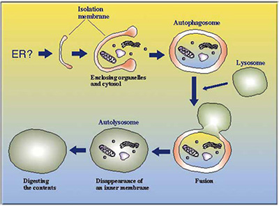
The process of autophagy
- Autophagy depends on large-scale membrane reconfigurations within the cell.
1.Formation of the autophagosome
- Membranes within the cell form a cup-shaped structure that expands around cellular material or organelles, eventually forming a double- (or multi-) membranous vesicle known as the autophagosome that isolates targets of degradation.
2.Fusion with the endosomes or the lysosome/vacuole
- The outer membrane of the autophagosome fuses with the membranes of endosomes or the lysosome/vacuole.
3.Degradation of the inner membrane of the autophagosome
- In the case of yeast, the inner membrane of the autophagosome is next released into the inside of the vacuole. This inner membrane of the autophagosome, now known as the autophagic body is subsequently degraded by proteases resident within the vacuole.
4.Degradation of cellular material
- Cellular material engulfed by the autophagosome in step 1, which is no longer protected by the membrane of the autophagic body, is finally degraded by lysosomal/vacuolar proteases.
Unanswered questions in autophagy research
- We are interested in the following areas:
1.The mechanism of autophagy induction
2.The mechanism of autophagosome formation, and the origin of lipids recruited to form the autophagosome
3.The link between autophagy and other forms of lysosomal and vacuolar transport pathways
4.The degradation mechanism of autophagic bodies
5.The physiological significance of autophagy in multicellular organisms
Our approach to research
Experimental materials
- Over many years of research, we have used yeast cells, animal cells, mice and Arabidopsis thaliana in our work.
Research methods
- Molecular genetic techniques - We isolate and clone autophagy-related genes, and incorporate into this process screening for genetic interactions, analysis of autophagy phenotypes in mutant cells, cloning of homologous genes and the generation of cells with no autophagic function.
- Molecular biological techniques - We employ the two-hybrid method to screen for physical interactions with autophagy proteins, analyse the regulation of gene expression and search for functional protein domains.
- Biochemical analysis - We interrogate the features of autophagy-related molecules through biochemical techniques. These methods include the generation of relevant antibodies, epitope tagging and the fractionation and isolation of organelles.
- Morphological analysis - We examine cellular morphology by light microscopy, employing techniques including immunofluorescence, examination of molecular localisation through the expression of proteins fused with GFP and electron microscopy
- Physiological analysis - We have established assays of autophagic activity and use these to uncover the conditions of autophagy induction as well as conditions that are able to suppress autophagy, and analyse the phenotypic consequences of autophagy-related gene deletion.
- Using these techniques, we aim to comprehensively describe the role of autophagy, from the perspective of both cell biology as well as the physiology of cells.
Conclusion
- For further information, please consult out published experimental work and review articles.
- T.Noda
Information
Copyright (C) 2016東工大大隅研究室 All Rights Reserved.